
How the Atlas (C-1) Misalignment Influences the Nervous System
Society of Brain Mapping and Therapeutics (SBMT)
Los Angeles, CA
March 15, 2024
Presented by Wontaek Hwang, DC
+ Head & Neck Spinal Care (2022)
+ NUCCA Wellness Clinic of Chicago (2018)
Notes:
We truly see in our everyday practices that the atlas (C-1) vertebra misalignment plays either a causative or contributory role in the nervous system dysfunction. There is special group of chiropractors called an upper cervical chiropractor. These people have been very disciplined to explore the connection between the atlas vertebra misalignment and its association with structural and neurological functions.
National Upper Cervical Chiropractic Association (NUCCA) has been developing this system of imaging protocols and analysis to deal with the small and measurable spinal misalignment in the craniocervical junction (CCJ) at the base of the skull. We call it, Atlas subluxation. This appears to be influencing lots of brain functions, and upper cervical chiropractors have been curious to understand how this small misalignment in the upper cervical spine can cause interference to the nervous system, and particularly, via brain vascular flow.
With gratitude to Scott Rosa, DC, Young H. Chung, DC, Ian Bulow, DC

Let’s take a look at the anatomy briefly.
The upper neck is the most complex joint in the spine. There are no spinal discs in between these joints. This joint relies on muscles and connective tissues to stabilize the brain and spinal cord. This atlas (C-1) needs to bear the weight of our skull while it provides the majority of the head rotation.
The nature of this structure of this joint makes this particularly vulnerable to injury. Sometimes, accidents, injuries, and repeated traumas can damage this joint in complex ways and can lead to instability.
When the atlas shifts out of its proper position, it’s no longer in alignment with the foramen magnum at base of the skull and superior articulating surface of C1, and this can lead to complications in the brain communication.

This region entails very important structures—such as brainstem, upper cervical spinal cord, brain vascular flow, CSF flow, etc.—that contribute significantly to our brain communications. So, the structural problem in this specific region of the head and neck can lead to something called, the Atlas Subluxation Complex Syndrome. Historically, chiropractic profession has been curious about how the spinal misalignments interfere with the nervous system. NUCCA chiropractors have been very interested to develop a system of analysis and corrective procedure to deal with the small and measurable misalignment in the craniocervical junctions called Atlas subluxation.

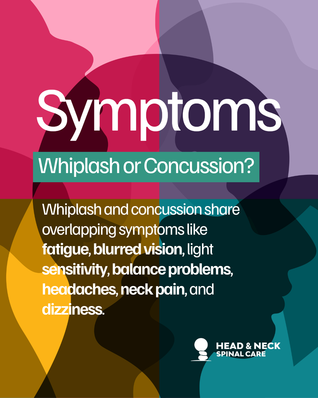
Public and professional recognitions have grown regarding how addressing this C1 misalignment can markedly reduce the severity and frequency of these signs and symptoms listed here.
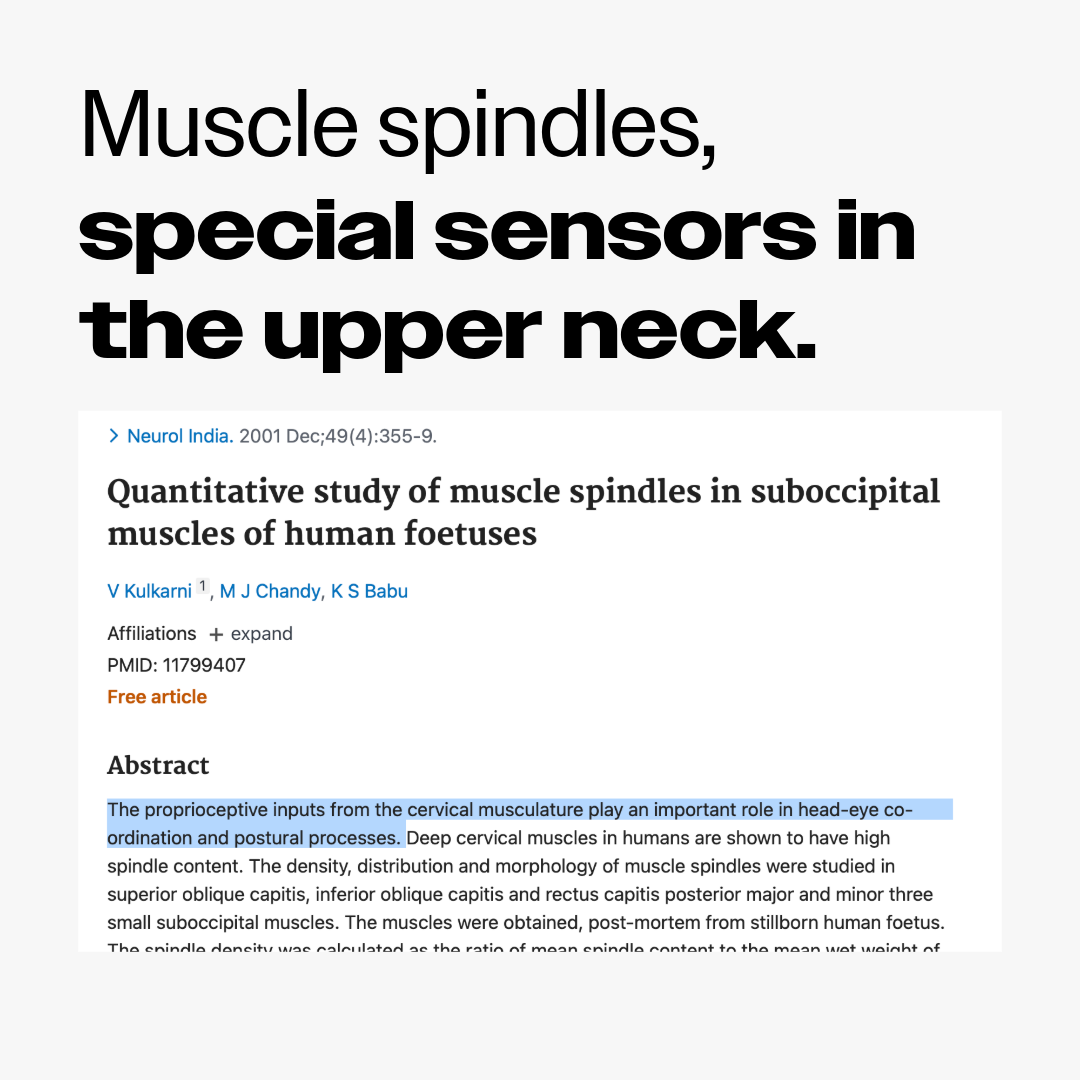
To us, it appears very clearly that the problem in our upper neck region may not only contribute, but in many cases, is the primary cause of many neurological disorders including autonomic nerve problems—such as breathing, heart beating, eye coordination, posture control, blood pressure, etc.

#1 Mechanical compression on the spinal cord
The concept of direct mechanical tension on the spinal cord, it seems quite straightforward, but it’s still unclear.
We know, practically speaking, that there’s quite a direct correlation between the atlas subluxation and spinal cord functions. After a proper adjustment, it’s not rare to see improvements or changes in the lower extremities such as sciatic pain or sensation of warmth, and even swelling.
However, the physical compression on the spinal cord in the upper cervical junction does seem very challenging because of the large gap between the cord and its spinal canal wall.
Dr. Richard Grostic, a NUCCA chiropractor, came up with the dentate ligament hypothesis, that perhaps, this short and thick ligament might be responsible for changes in spinal cord functions.
*If you’d like to know more about this, please visit the link below. Well-written article by Erikson, K.
https://musculoskeletalkey.com/neurophysiology-and-the-upper-cervical-subluxation/

There’s this single fibrous strip, laterally projecting ligament, very strong ligaments that anchors the spinal cord against motion.
In the upper cervical spine area, this ligament pass perpendicular to the cord, he suggests that this ligament has a restraining function from the vertical movement.
This cross section of the spinal cord shows that the nerve tracts like spinothalamic or spinocerebellar tracts lie very close to the dentate ligament attachment points. We suspect that the traction on this strong ligament due to the atlas subluxation can stress and deform the spinal cord.

#2 Obstruction in brain venous outflow
It’s been also known that anatomical position of atlas vertebra can influence on changes in circulation of vertebral artery. We also suspect that the structural misalignment in the craniocervical junction would influence on the brain vascular flow.
According to Dr. Richard Grostic, he claims that it’s been observed that the small radicular veins of upper cervical spinal cord were not as redundant as elsewhere in the spine. Mechanical obstruction of these veins could cause disruption to brain venous outflow. Because these veins run at low pressure, he suspects that the veins can easily be influenced by, and vulnerable to, physical compression.


The cerebrospinal fluid flow (CSF flow) plays a crucial role in brain vascular system as well. CSF flows over the surface of the brain and down the spinal cord in the subarachnoid space, providing protection and removing metabolic waste from the brain. The CSF flow is thought to be driven by arterial pulsations and is closely linked to the cardiac cycle. This relationship suggests a deeper connection between CSF flow and arterial dynamics.
More and more studies are suggesting that cerebrospinal fluid dynamics involves with increased pressure in our brain, and such brain fluid imbalance can lead to secondary neurodegenerative disorders. Dr. Scott Rosa, the same upper cervical chiropractor, did utilize special MRI imaging to visualize CSF flow in his patient with cerebellar tonsilar ectopia. He found out that there’s pooling of CSF in the frontal part of the brain in his patient. He was able to see improvements in CSF flow before and after followed by proper correction in the upper cervical spine.

Marshall Dickholtz, Sr. published a study about the association between the atlas alignment and arterial blood pressure.
Dr. Dickholtz, Sr. was a NUCCA chiropractor, and this interview led me to become familiar with the idea of our neck affecting the whole body. He published a pilot study with Dr. Bell who was a family physician based in Barrington, Illinois, they concluded that there’s an association between the atlas alignment and reduction in arterial blood pressure for sustained period of time.
So, the idea is not to promote that correcting this atlas subluxation would reduce the blood pressure, but to share a perspective that the structural problem in this craniocervical junction can contribute to our whole body system.

Utilization of CBCT Scan in Upper Cervical Practices
The clinical use of this CBCT has been present over 20 years, and it’s been mostly used in dentistry. Now, they are being introduced to upper cervical chiropractic offices as well. This advanced imaging has allowed us to reduce errors in patient positioning, and to visualize in 3D high quality images to understand the underlying problems or anatomical abnormalities for less or equal radiation compared to traditional X-ray radiographs.
Workcited
Bakris G, Dickholtz M Sr, Meyer PM, Kravitz G, Avery E, Miller M, Brown J, Woodfield C, Bell B. Atlas vertebra realignment and achievement of arterial pressure goal in hypertensive patients: a pilot study. J Hum Hypertens. 2007 May;21(5):347-52. doi: 10.1038/sj.jhh.1002133. Epub 2007 Mar 2. PMID: 17252032.
Bothwell, S. W., Janigro, D., & Patabendige, A. (2019). Cerebrospinal fluid dynamics and intracranial pressure elevation in neurological diseases. Fluids and Barriers of the CNS, 16(1). https://doi.org/10.1186/s12987-019-0129-6
DeNunzio G, Evans T, Beebe ME, Browning J, Koivisto J. Craniocervical Junction Visualization and Radiation Dose Consideration Utilizing Cone Beam Computed Tomography for Upper Cervical Chiropractic Clinical Application a Literature Review. Dose Response. 2022 Jun 13;20(2):15593258221107515. doi: 10.1177/15593258221107515. PMID: 35719850; PMCID: PMC9201332.
Erikson, K. (2016), Figure 15-1, Figure 15-5A, Figure 15-6, Neurophysiology and the Upper Cervical Subluxation. , Modified from Eriksen K. Upper Cervical Subluxation Complex. Baltimore: Lippincott Williams & Wilkins, 2004:75. https://musculoskeletalkey.com/neurophysiology-and-the-upper-cervical-subluxation/
Flanagan, Michael F. “The Role of the Craniocervical Junction in Craniospinal Hydrodynamics and Neurodegenerative Conditions.” *Neurology Research International*, vol. 2015, 2015, pp. 1–20, https://doi.org/10.1155/2015/794829.
Mestre, Humberto, et al. “Flow of Cerebrospinal Fluid Is Driven by Arterial Pulsations and Is Reduced in Hypertension.” *Nature Communications*, vol. 9, no. 1, 19 Nov. 2018, https://doi.org/10.1038/s41467-018-07318-3.
Seragioli, Rafael et al. “Assessment of the cervical spine denticulate ligament using MRI volumetric sequence: Comparison between 1.5 Tesla and 3.0 Tesla.” Journal of neuroradiology. Journal de neuroradiologie 45 2 (2017): 147-151
Rosa, S., Baird, J. W., Harshfield, D., & Chehrenama, M. (2018). Craniocervical Junction Syndrome: Anatomy of the Craniocervical and Atlantoaxial Junctions and the Effect of Misalignment on Cerebrospinal Fluid Flow. InTech. doi: 10.5772/intechopen.72890